Home
Atlas of Genitourinary Pathology
Barnes and Noble
Loading Inventory...
Atlas of Genitourinary Pathology
Current price: $119.99
Barnes and Noble
Atlas of Genitourinary Pathology
Current price: $119.99
Loading Inventory...
Size: OS
*Product information may vary - to confirm product availability, pricing, shipping and return information please contact Barnes and Noble
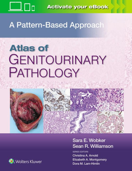
Atlas of Genitourinary Pathology
provides a single referral source for images and information concerning all pathological entities in the field of genitourinary pathology. The book contains gross photos and photomicrographs of pathologic entities, and variants of those entities, occurring in the following organs or anatomic sites:
• Adrenal
• Kidney
• Renal pelvis and ureter
• Urinary bladder
• Urethra
• Prostate
• Seminal vesicles
• Testis
• Spermatic cord and testicular adnexae
• Penis and scrotum
The book is lavishly illustrated with images accompanied by text explaining the key diagnostic points, as well as features that help separate the entity from similar lesions. Images from the book are available in powerpoint format from the Extra Materials website.
A comprehensive and authoritative work for practicing pathologists and pathologists in training as well as medical students, urologists, radiation oncologists, and medical oncologists.