Home
Histology: An Interactive Virtual Microscope / Edition 1
Barnes and Noble
Histology: An Interactive Virtual Microscope / Edition 1
Current price: $61.95
Barnes and Noble
Histology: An Interactive Virtual Microscope / Edition 1
Current price: $61.95
Size: OS
Loading Inventory...
*Product information may vary - to confirm product availability, pricing, shipping and return information please contact Barnes and Noble
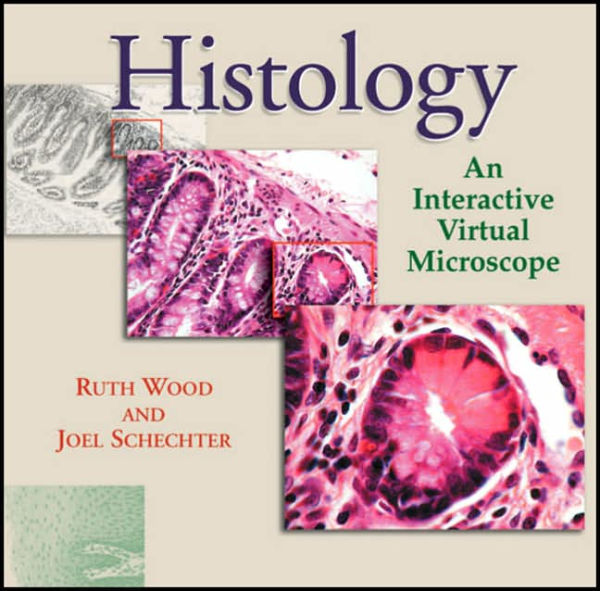
Histology: An Interactive Virtual Microscope
recreates the look and feel of a microscope in an intuitive, browser-based interface. Histology is one of the core courses in the curriculum for medicine and allied health professions. It requires both an understanding of the cellular structure of organs and tissues and the ability to recognize tissues at the microscopic level. Traditionally, students have used microscopes to study specimens on glass slides. While extensive practice with the microscope is not essential in the modern health care setting, histology remains an image-intensive discipline. To understand the organization and function of tissues requires that students be able to interpret microscopic images. By presenting nested images at increasing magnification,
provides a sense of scale and proportion that cannot be achieved in a standard histology text or atlas. Moreover, the text descriptions and labeled images offer flexibility (students can study at any computer) and opportunities for small-group learning (several students can examine the images together), yet provide guidance for independent study.